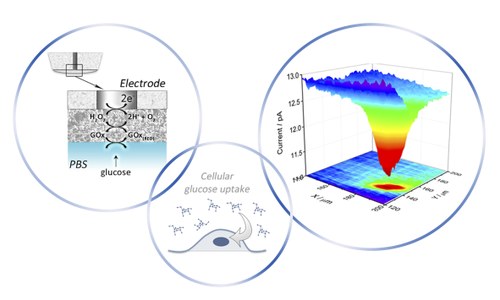
Tumor cells show altered oxygen consumption, an impaired glucose consumption and consequent release of lactic acid. Rapino’s group has investigated the oxygen consumption in cell cultured in adhesion and in sospension, the glucose consumption and lactate release in living cells using microsensors as probes of SECM. This tool allows the elucidation, at the single cell level, of the metabolic alterations that characterize the initiation and the development of cancer.
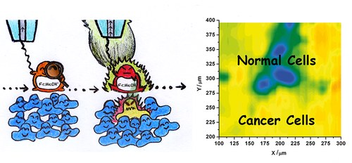
Cancer cells show an imbalance in the ratio of oxidizing and reducing species. This imbalance could be used for diagnostic purposes. Dr. Rapino and collaborators has developed a strategy that uses SECM and ferrocenemethanol as redox molecule to explore and characterize the intracellular redox balance and particularly the increased reduced glutathione levels in cancer cells. The recorded SECM maps show different feedback currents for tumor and normal cells. Both culture cells and patient cells were explored.
[Electrochimica Acta 2015; 179:65-73]
