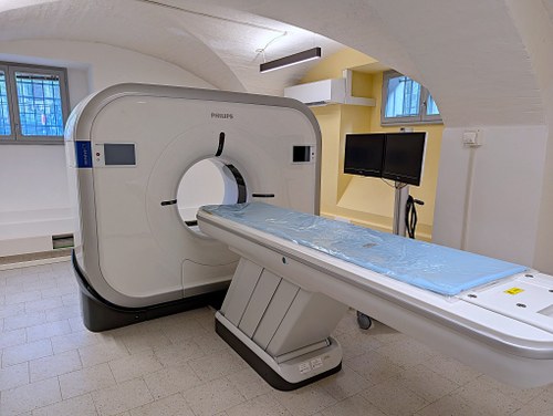
CT, which stands for Computed Tomography, is a diagnostic technique that uses ionizing radiation (or X-rays) to obtain detailed images, in a three-dimensional version, of specific anatomical areas of the human body (e.g., brain, bones, blood vessels, abdominal organs, thoracic organs, respiratory tract, etc.). In particular, the machine present at our Center uses a technique called helical (or spiral) computed tomography in which a continuous movement of the couch through the gantry is exploited, while the X-ray tube rotates around the patient: this generates a helical diagnostic pattern. This modality allows volumetric images to be acquired in a shorter time than conventional CT, improving image quality and reducing the risk of motion artifacts. It is particularly useful for examining internal organs, blood vessels, and soft tissues, and is used in the medical field for diagnosis of cardiovascular, oncological, and neurological diseases.
The one present in our Center is the first CT for research purposes in Italy.
For service use of the instrument and/or possible scientific collaborations, please make contact with:
TECHNICAL CHARACTERISTICS:

