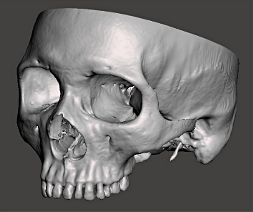
Clinical-anatomy research topics are focused on evaluation of anatomical variants and related clinical implications, developing of new surgical techniques in head and neck region in particular in oral-facial district, carried out on embalming and fresh donor bodies.
Facial skeleton plays an important role in the appearance of the face and in its many functional aspects. It represents the structural support of the face and its muscles, influencing chewing, speech, swallowing and the function of the upper airways.
Anatomy Center studies are focused on the development of surgical techniques for the use of grafts of autogenous pedicled tissue, free flaps of soft and hard tissues, such as the pericranial galea flap, pedicled or free, considered a goal strategic operational choice for maxillofacial surgeons. The design of this type of flap includes two layers of fascia and periosteum with osteogenic potential taken from the temporal region, including its superficial vascular network, creating an anastomosis between the superficial temporal and cervical vessels to restore the newly regenerated tissues of the recipient site. Thanks to the new computer-assisted technologies used in the study, such as augmented reality (AR), specific surgical endpoints are evaluated to be developed and allow direct observation of the patient's anatomy and the surgical field, facilitating anatomical visualization during the collection of the free flap.
Other studies are focused on the analysis of the structural changes of the orofacial district and pharyngeal volume, following the use of advanced intraoral devices on donor boby, studying the obstructive sleep apnea syndrome (OSAS) by using Computed Tomography (CT).