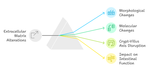
Full-thickness tissue analyses in patients with severe dysmotility (SD), both idiopathic and genetic, have revealed extracellular matrix alterations at both morphological and molecular levels.
It has long been known that the crypt-villus axis and the extracellular matrix component play a crucial role in the proper differentiation of intestinal mucosal cells. The extracellular matrix not only provides structural support but also acts as a dynamic platform regulating numerous biological processes, including cell proliferation, migration, and differentiation.
In the crypt-villus axis, the interaction between crypt stem cells and the extracellular matrix is essential for maintaining the balance between cell renewal and epithelial maturation. Alterations in the matrix can disrupt this delicate equilibrium, interfering with proper cellular turnover and causing dysfunctions in the intestinal barrier.
When the regulatory program of this axis fails to function properly, the consequences can be dramatic, significantly impacting nutrient absorption and intestinal homeostasis.
A detailed analysis of the composition, structure, and collagen formation process in SD-affected tissues represents an opportunity to further explore the functional aspects of the disease. Understanding how matrix alterations influence the physiology of the crypt-villus axis can not only shed light on pathogenic mechanisms but also help identify potential therapeutic targets to improve the management of this complex condition.

